
Page 95 - Slipped Capital Femoral Epiphysis Pathogenetic and Clinical aspects
P. 95
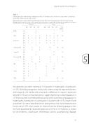
Table 3
Semiquantitative immunohistochemistry profiles of chondrocytes and bone components (osteoblasts, - clasts and - cytes) in cases and controls.
CC chondrocyte cytoplasm, CN chondrocyte nucleus, OC osteocyte cytoplasm, ON osteocyte nucleus
GHR Growth hormone receptor, TR α and β Thyroid receptor α and β, IGF insuline like growthfactor receptor
Caspase 0,0
0,5 1
Androgen R Leptin R Estrogen R β
IGF
3 0
0
16 16 1 1
3 0,5 0
3 0 1 2
8 15 7 16
92 17 1 13 13 13 9 44 3 7
20 20
2
1,0
0,0
0,5
0,0
0,5211
0,0 3 3 8 7 0,5 0 0 5 9 1,0 0 0 4 1
0,0 2 0
0,53 4
1,013 110
Slipped Capital Femoral Epiphysis
Control
Case
CC
CN
OC
ON
CC
CN
OC
ON
max TOTAL
GHR 0,5 1,0 TRα 0,0
4
5 1 3 2 0 1 1 2 1 1
3 0
1
1 18 8
S1000,00 1
0,5 4
1,0 6 TRβ0,0
0,5
2 17
5 0
1 1 15 6
24
30
1825 2
20 5 19 20
5
We observed consistent staining of S100 protein in hypertrophic chondrocytes in SCFE, facilitating recognition and actually underscoring the representativeness of the biopsies [20]. Neither did we observe a difference in Caspase 3 expression between SCFE cases and controls physes, suggesting that no increased apoptosis in SCFE had occurred. Our finding did not concur with a study measuring apoptosis in hypertrophic chondrocytes in core biopsies in a patient with SCFE using the Tunel procedure [1]. It seems therefore that this phenomenon may not be representative for all cases of SCFE, since Caspase 3 is more sensitive for detecting apoptosis than the Tunel procedure [8]. Increased expression of CD34 in SCFE physes, as marker for vessel density, would point inflammatory activity accompanying slippage.
93
