
Page 94 - Slipped Capital Femoral Epiphysis Pathogenetic and Clinical aspects
P. 94
Chapter 5
receptor alpha (SP1) showed no expression in either SCFE cases or controls. All of the receptors did not show any statistical differences in both groups, SCFE and the controls (See table 3). We also correlated the differences in age (< or ≥11 years old), differences in sex or BMI (< or ≥ 25 kg/m2) with staining of the biopsies but found no significant relationships (data not shown). The groups acute, acute on chronic or chronic as well as stable/ unstable SCFE were too small for meaningful comparison.
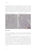
Figure 4A control IGFR1 staining (obj. 5x) Figure 4B SCFE IGFR1 staining (obj. 5x)
Discussion
The cause and pathogenesis of SCFE remains obscure. We have studied the histomorphology of slipped physes in symptomatic patients and compared them to control physes.
The most obvious histological difference in SCFE compared with controls was consistent perturbation of the linear architecture of hypertrophic chondrocytes, a finding similar to that of other studies [2, 3, 9, 11]. The loss of longitudinal orientation of the chondrocytes in SCFE physes was probably due to tangential forces during slippage in SCFE [7]. Our findings of irregular columns are largely in line with earlier reports in the literature, as shown in table 4, but we could not confirm diminished numbers of chondrocyte [2, 10, 11, 15]. The resting zone of the epiphyses with SCFE was located outside the slipping zone and appeared to be relatively normal [2, 10, 15].
92
