
Page 93 - Slipped Capital Femoral Epiphysis Pathogenetic and Clinical aspects
P. 93
Immunohistochemistry
CD34 and Caspase activity
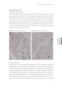
SCFE and controls CD34 staining was observed in the transition zone of the physis towards the metaphysis, while no signs of neovessels were seen around hypertrophic chondrocytes. In 9 SCFE cases, we did not observe signs of (increased) vascularity, while in 9 SCFE cases limited signs of increased vessels size and number were seen. For the control group there was positive vessel staining in 3 out of 10 biopsies (See figure 3A and B). No statistical differences between SCFE and the controls were noted. The expression of Caspase-3 was consistently faint to absent in all cases and controls (not shown).
Figure 3A Control CD34 staining (obj. 5x) Figure 3B SCFE CD34 staining (obj. 5x)
5
Slipped Capital Femoral Epiphysis
Hormonal receptors
Expression of Insulin-like growth factor receptor (IGFR1) and growth hormone receptor expression was observed in the cytoplasm of chondrocytes and osteocytes, but showed no statistically significance between cases and controls (see figure 4A and B). Estrogen β (ER-β) receptor stained only the nucleus of both the osteocyte and chondrocyte, but showed no statistically significance between cases and controls. Thyroid receptor α was faintly expressed in cytoplasm and nucleus of the chondrocyte and osteocyte, in contrast with Thyroid receptor β, which was consistently not detected. Androgen receptor, leptin receptor and estrogen
91
