
Page 54 - PIECES OF THE PUZZLE Eline Vissia
P. 54
CHAPTER 3
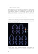
Hippocampal shape analysis
As compared with HC, All-PTSD (Figure 3.1b) as well as both PTSD-DID (Figure 3.1c) and PTSD-only (Figure 3.1e) showed deformations in the CA1, CA2-3 and subiculum. Direct comparison of shape measures between PTSD-DID and PTSD-only showed relative contractions in the CA1, CA2-3 and subiculum in PTSD-DID (Figure 3.1d). The results of these shape analyses did not survive multiple comparison correction with permutations. Uncorrected shape deformation results are presented for exploratory purposes because they support and inform on the significant volumetric results.
Figure 3.1 a) A schematic representation of the hippocampal subfields mapped onto a representative hippocampal surface obtained by averaging the surface from all the participants. In addition, 3D maps of regional hippocampal shape differences (uncorrected) are shown comparing (b) All-PTSD vs. HC, (c) PTSD-DID vs. HC, (d) PTSD-DID vs. PTSD-only and (e) PTSD-only vs. HC. Upper rows represent anterior view and lower rows represent posterior view.
Abbreviations:
PTSD-only = patients with only posttraumatic stress disorder; PTSD-DID= patients with PTSD and dissociative identity disorder; All-PTSD= includes both PTSD-only and PTSD-DID patient groups; HC= healthy controls.
116
