
Page 60 - Cardiac abnormalities after aneurysmal subarachnoid hemorrhage
P. 60
Chapter 4
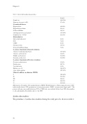
Table 1: shows the baseline characteristics.
Female sex
Mean age (in years)( ±SD)
Coronary risk factors
Hypertension
Hypercholesterolemia
Diabetes Mellitus
Smoking (previous and current)
Family history of CVD
Medical history
Myocardial infarction*
PCI
CABG
Ischemic Stroke
Intracerebral Hemorrhage
Location of aneurysm in Carotid circulation Anterior Communicating Artery
Anterior cerebral artery
Middle cerebral artery
Internal carotid artery
Location of aneurysm in Posterior circulation Posterior cerebral artery
Basilar artery
Vertebral artery
Hijdra score >19
Acute hydrocephalus
Clinical condition on admission (WFNS)
I
II
III
IV
V
210(70%) 57(±13)
90(30%) 26(9%) 14(5%) 115(38%) 33(11%)
8(3%) 7(2%) 4(1%) 16(5%) 7(2%)
127(42%) 12(4%) 61(20%) 61(20%)
11(4%) 20(7%) 9(3%) 142(47%) 107(35%)
103(34%) 54(18%) 24(8%) 64(21%) 56(19%)
N=301
Abbreviations: N: number, SD: standard deviation, WFNS: World Federation of Neurosurgical Societies. CVD: cardiovascular disease. PCI: percutaneous coronary intervention, CABG: coronary artery bypass graft *: The patients with prior myocardial infarction, PCI or CABG who were included did not have ECG abnormalities or echocardiographic abnormalities prior to the SAH.
Cardiac abnormalities:
The prevalence of cardiac abnormalities during the study period is shown in table 2.
58
