
Page 67 - Slipped Capital Femoral Epiphysis Pathogenetic and Clinical aspects
P. 67
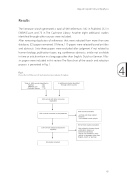
Results
The literature search generated a total of 689 references: 382 in PubMed, 232 in
EMBASE.com and 75 in The Cochrane Library. Another eight additional studies
identified through other sources were included.
After removing duplicates of references that were selected from more than one
database, 525 papers remained. Of these, 119 papers were selected based on titles
and abstracts. Sixty-three papers were excluded after judgment if not related to
human biology, publication types, e.g. conference abstracts, article not available
in time or article written in a language other than English, Dutch or German. Fifty-
six papers were included in this review. The flow chart of the search and selection
process is presented in Fig. 1. 4
Fig. 1.
Flow chart of the search and selection procedure of studies
J Child Orthop
Fig. 1 Flow chart of the search and selection procedure of studies
Chronic disease: reviews the effects of chronic disorders on the physis
Diagnostic endocrine measurements in SCFE: reviews the studies that specifically investigated SCFE
The physis in SCFE
Histological changes in SCFE
Histological studies of tissue obtained from biopsies during
The proliferative and hypertrophic zones of the epiphysis in SCFE were widened compared with normal physis, and showed irregular columnar organisation with gradual loss of longitudinal septa and diminished number of chondrocytes in each column [3–7]. Interestingly, Adamczyk et al. showed that apoptosis was increased throughout the physis in SCFE, in contrast with controls, where apop6t5osis was found only in the hypertrophic zone [8]. The chondrocytes showed intracellular abnormalities [5, 7, 9]. An increase in the nuclear and cytoplasmic density was seen in the pro-
Slipped Capital Femoral Epiphysis
