
Page 106 - Slipped Capital Femoral Epiphysis Pathogenetic and Clinical aspects
P. 106
Chapter 6
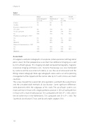
Table 1.
Continued
K-wires 3% Variable 2% Capsular compression (n = 133)
Yes–open 4% Yes–by aspiration 28% No 68% Prophylactic pinning (n = 135)*
Yes 6% No 9% Depends 85% Removal of metalwork (n = 135)*
Yes 17% No 34% Depends 49%
Assessment
3% 3% 0% 1%
13% 6% 10% 10% 23% 26% 77% 71% 65%
15% 9% 12% 36% 17% 88% 49% 74%
33% 21% 12% 39% 36% 88% 28% 43%
BSCOS
WKO
POSNA
United Kingdom (n = 149)
Netherlands (n = 71)
Total
(n = 220)
n = 794
All surgeons used plain radiographs in two planes (antero-posterior and frog-lateral pelvic views) for their preoperative assessment but additional imaging was used by 30% of both groups. This imaging included computed tomography, magnetic resonance imaging and bone scans. Dynamic fluoroscopy was also mentioned by some to aid the assessment of stability as was the use of ultrasound and the Billings lateral radiograph. Bone age radiographs were used as an aid to planning management of the slipped and the normal sides by 6.7% with similar use in both countries.
The survey showed that, overall, 68% of respondents used both the acute/chronic and the unstable/stable methods of classification. Some significant differences were observed within the subgroups of this study. The use of both systems was more common in those with a larger paediatric practice (> 50% of workload) than in those with a more mixed practice, 74% compared with 58% (P < 0.05); and in the UK rather than in the Netherlands, 75% compared with 51% (P < 0.05). The Southwick classification [7] was used by only eight surgeons (5%).
104
