
Page 63 - Cardiac abnormalities after aneurysmal subarachnoid hemorrhage
P. 63
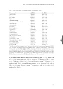
Time course and risk factors for myocardial dysfunction after aSAH Table 3: shows the univariable ORs with determinants for early and late WMA.
Determinants*
Early WMA
Late WMA
Age > 50 years
Female sex
Poor WFNS
Hijdra >19
Acute hydrocephalus
Aneurysm in carotid circulation† Sinusbradycardia Sinustachycardia
Low voltage
Pathologic Q wave
ST depression
ST elevation
Negative T-waves
Prominent U-waves
QTc prolongation > 500ms LVH on ECG
Strain pattern on ECG Myocardial infarction pattern Elevated Troponin T Elevated NT-proBNP Decreased GFR
1.7(0.9-3.4) 0.6(0.3-1.2) 3.0(1.6-5.5)‡ 1.4(0.8-2.4) 0.6(0.3-1.1) 1.1(0.6-2.1) 0.4(0.1-1.3) 3.3(1.3-8.9)‡ 3.2(0.8-11.8) 1.0(0.99-1.0) 5.8(2.4-14.4)‡ 31.8(3.8-264.5)‡ 2.4(1.3-4.8)‡ 0.6(0.3-1.1) 1.7(0.8-3.7) 0.7(0.5-1.1) 0.8(0.2-3.7) 3.6(1.0-12.1)‡ 4.2(2.2-7.9)‡ 2.6(1.04-6.7)‡ 0.96(0.9-1.0)
0.6(0.3-1.5)
0.9(0.4-2.2)
1.1(0.5-2.6)
0.9(0.4-2.2)
0.5(0.2-1.1)
1.9(0.8-4.4)
0.5(0.1-2.4)
2.9(0.9-9.9)
9.6(1.8-50.7) ‡
1.0(0.99-1.0)
2.5(0.7-9.8)
NE
1.5(0.6-3.6)
1.1(0.5-2.7)
1.8(0.6-5.3)
0.8(0.5-1.2)
0.9(0.1-7.8) 4 8.0(2.0-32.2)‡
3.9(1.6-9.7)‡ 1.6(0.5-5.2) 0.99(0.9-1.1)
*The ORs of medical history (angina pectoris, myocardial infarction, prior coronary artery bypass graft(CABG), prior angioplasty, known hypertension, hypercholesterolemia, peripheral vascular disease, cerebrovascular events(ischemic or hemorrhagic), diabetes mellitus, current cigarette smoking or family history of coronary artery disease) with early and late WMA are not shown as they were not significantly associated. † includes anterior communicating artery, anterior cerebral artery, middle cerebral artery and internal carotid artery. ‡ These determinants were included in the multivariable analyses. Abbreviations: LVH: Left ventricular hypertrophy; WMA: wall motion abnormalities; GFR: glomerular filtration rate. NE: not estimable.
In the multivariable analyses (determinants marked in table 3), poor WFNS OR 2.7 (1.1-6.8), sinus tachycardia OR 5.0 (1.3-19.9), ST-depression OR 3.7 (1.02- 13.1), ST-elevation OR 16.6 (1.5-178.9) and elevated troponin T OR 2.8 (1.1-7.3) predicted early WMA. Myocardial infarct pattern on the admission ECG with an OR 6.8(1.6-30) and elevated troponin T on admission with an OR 3.4 (1.4-8.5) predicted late WMA.
61
