
Page 71 - Slipped Capital Femoral Epiphysis Pathogenetic and Clinical aspects
P. 71
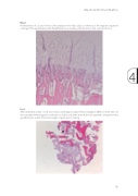
Fig. 2.
Normal physis of a 2-year-old boy after amputation for tibial aplasia. At the top is the regularly organized cartilage of the growth plate, with the different zones leading at the bottom to the ossification zone
Fig. 3.
Abnormal physis taken of a 10-year-old boy with slipped capital femoral epiphysis (SCFE) on both sides. At the top-right of the image, the ossification is visible, and at the bottom, the disorganized cartilage from the growth plate is visible. The normal regular organisation is lacking
4
Slipped Capital Femoral Epiphysis
69
