
Page 117 - Slipped Capital Femoral Epiphysis Pathogenetic and Clinical aspects
P. 117
was changed in three directions relative to the acetabulum: flexion, varus and derotation. Peroperative fluoroscopy was performed to verify the position of the seating chisel and screws. All patients with an unilateral slipped hip were treated with a preventive K-wire fixation at the contralateral hip. Patients were not allowed to bear weight for 6 weeks.
Table 1.
Patient data
Gender ratio of patient cohort, n M: 16, F: 12 Mean age at surgery, years 13 Unilateral (left/right), n 15 Bilateral, n 13
ITO unilateral (left/right), n 24 ITO bilateral, n 4 Acute on chronic, n 5 Chronic (hips), n 27 Trauma in history, n 13
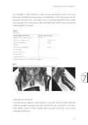
ITO intertrochanteric osteotomy; M male; F female Fig. 2.
(range 9–17) (10/5)
(15/9)
One-year postoperative SCFE after epiphysiodesis and Imhauser osteotomy
7
Slipped Capital Femoral Epiphysis
Demographic/clinical data
Patient cohort (n = 28)
Outcome assessments
Data are based on patients’ notes and the X-ray results. Patients were traced and asked to complete questionnaires [part of the Harris Hip Score (HHS) and Short- Form Health Survey (SF-36)] at home; they also were invited for a clinical and radiological evaluation.
115
