
Page 46 - ADD-ON ABLATION SURGERY IN PATIENTS WITH ATRIAL FIBRILLATION
P. 46
Chapter 2
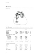
Figure 1: box lesions
Posterior view of the heart. The bold lines illustrate the six epicardial ablation lesions, encircling all four
pulmonary veins.
Table 1. Comparison of baseline characteristics and rhythm outcome between control and add-on surgery patients (N=132).
Study Population
Control (N=67)
Add-on surgery (N=65)
p-value
Demographic data
Age (Years) Weight (Kg) Sex (male)
68.2 ± 9.1 77.9 ± 16.4 85 (64.4%)
71.0 (38.8 – 85.0) 77.7 (50 – 170) 39 (58.2%)
30(44.8%) 21(31.3%) 15(22.4%) 1(1.5%)
84,1 (3 – 618) 50.4 (33-70) 56.5 (30-80)
34 (44.7%) 61 (80.3%) 33 (44%) 3 (3.9%) 23 (30.3%) 41 (53.9%) 7 (9.2%)
61.9 (46.6 – 81.0) .17 78.1 (49 – 111) .89 46 (70.8%) .13
.99
27(41.5%)
22(33.8%)
15(23.1%)
1(1.5%)
78,0 (33 – 403) .73 50.7 (40-67) .38 48.8 (18-79) .01
24 (33.3%) .16 59 (81.9%) .79 27 (37.5%) .42 4 (5.6%) .65 18 (25%) .47 35 (48.6%) .52 5 (7.0%) .63
Previous cardiac history (N=132)
Atrial Fibrillation: Paroxysmal AF Permanent AF
Persistent AF
Atrial flutter Total months of AF
Left Atrial Dimension (mm) LeftVentrical Ejection Fraction (%)
57(43.2%) 43 (32.6%) 30 (22.7%) 2 (1.5%)
81.0 ± 102.4 50.6 ± 7.5 52.6 ± 13.5
Pre-operative complaints (N=132)
Palpitations Dyspnea Angina
(Pre-) Syncope Dizziness Fatigue
Other complaints
58 (39.2%) 120 (81.1%) 60 (40.8%) 7 (4.7%)
41 (27.7%) 76 (51.4%) 12 (8.2%)
54
