
Page 55 - Maximizing the efficacy of ankle foot orthoses in children with cerebral palsy
P. 55
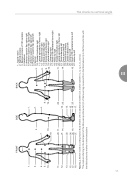
The shank-to-vertical angle
55
III
1. 2.
3.
1. 2. 4.
3.
4.
7.
6.
5. 10.
19.
10.
8. 19.
9. 10.
11. 12.
20. 11.
20.
11.
13.
13.
15. 17. 18.
24.
26. 16. 27.
15.
18. 25.
16.
FRONT
SIDE
BACK
1. Jugular notch
2. Xyphoid process
3. Spinous process of 10th vertebra 4. Umbilicus
5. Anterior superior iliac spine right
6. Posterior superior iliac spine right 7. Anterior superior iliac spine left
8. Posterior superior iliac spine left
9. Sacrum
10. Greater trochanter right
11. Lateral epicondyle of femur right 12. Tibial tuberosity right
13. Distal point tibia *right
14. Dorsal shell orthosis right
15. Lateral malleolus right
16. Calcaneus right
17. Head of 5th metatarsal bone right 18. Hallux tip right
19. Greater trochanter right
20. Lateral epicondyle of femur left 21. Tibial turberosity left
22. Distal point tibia* left
23. Dorsal shell orthosis
24. Lateral malleolus left
25. Calcaneus left
26. Head of 5th metatarsal bone left 27. Hallux tip left
21. 14. 22.
12. 23.
14.
17.
Figure 3.2. Marker model according Human Body Model, with six additional markers (i.e. marker numbers 12, 13, 14, 21, 22, and 23).
*The markers referring to the distal point of the tibia were positioned at 75% of the lower leg, measured from the tibial tuberosity to the floor and in line with the tibial tuberosity marker in the frontal plane
