
Page 96 - scheppingen
P. 96
four
94
dysplastic regions where prominent gliosis and presence of dysmorphic neurons and balloon or giant cells (in FCD IIb and TSC, respectively) was observed. Constitutive and particularly immunoproteasome subunits displayed increased expression compared to control, but also compared to mMCD specimens from patients with chronic epilepsy. These results indicate that increased expression of proteasome subunits is not simply an effect of seizure activity; moreover, the duration of epilepsy in mMCD cases did not
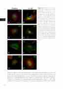
Figure 6 Effects of IL-1β stim- ulation on proteasome subunit expression in astrocytes in cell culture. Expression of β1 (A and B; green), β1i (C and D; green), β5 (E and F; red) and β5i (G and H; green) in unstimulated human fe- tal astrocytes (left panels) and in astrocytes after exposure to IL-1β (24 hours; 10 ng/ml, right panels); increased expression of all sub- units was observed. A transloca- tion of particularly the β1i and β5i subunits, shifting from cytoplasmic to perinuclear-nuclear expression following IL-1β treatment was ob- served. Cells were counterstained with phalloidin (actin filaments; red in A-D and G-H, green in E and F) and diamidino-2-phenylindole, DAPI (nuclei; blue). Scale bar in A: 15 μm.
