
Page 57 - scheppingen
P. 57
CODING AND SMALL NON-CODING TRANSCRIPTIONAL LANDSCAPE OF TSC
sine (20 μg/ml) and laminin (40 μg/ml) in 24-well plates at a density of 100,000 cells/ well. The cultures were grown in Neurobasal medium (NB) supplemented with B27, 0.35 mM HEPES, 200 mM L-glutamine, 14.3 mM β-mercaptoethanol and penicillin/strepto- mycin. Cultures were co-transfected at 1 day in vitro with miR34b-5p miRVana mimic (Applied Biosystems, Life Technologies Europe BV, Bleiswijk, Netherlands) or the miR-ID- IAN miRNA mimic negative control #1 (Dharmacon, GE Healthcare Europe, Eindhoven, the Netherlands) and a green fluorescent protein (GFP) vector at 50 pmol/well using Lipofectamine®-2000 (Thermo Fisher Scientific) as transfection reagent for 1 hour at 37oC and 5% CO2. Cultures were fixed with 4% paraformaldehyde/4% sucrose in phos- phate-buffered saline (PBS) at 4 days in vitro for 20 minutes and washed three times with PBS for 30 min at room temperature. The cultures were blocked with 0.1% PBS-Triton X-100 buffer with 3% Normal Goat serum and then incubated with primary antibodies in the blocking solution overnight at 4oC. Subsequently, neurons were washed three times
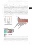
Figure 1 The transcriptome of tuberous sclerosis complex (TSC) cortical tubers determined by using RNA-Seq data (a) Volcano plot showing differential expression of genes between TSC tubers (n = 12) and post-mortem control cortex (n = 10). A total of 269 mRNAs were found to be over-expressed and 169 under-expressed in TSC tubers compared to control cortex tissue (b) Ingenuity pathway analysis showing major pathways enriched for over-expressed genes in TSC tubers (c) Heat map showing genes of the complement system enriched in TSC tubers compared to control cortex. All p-values are BH adjusted.
55
three
